

 16 hours ago
4
16 hours ago
4
PROTECT YOUR DNA WITH QUANTUM TECHNOLOGY
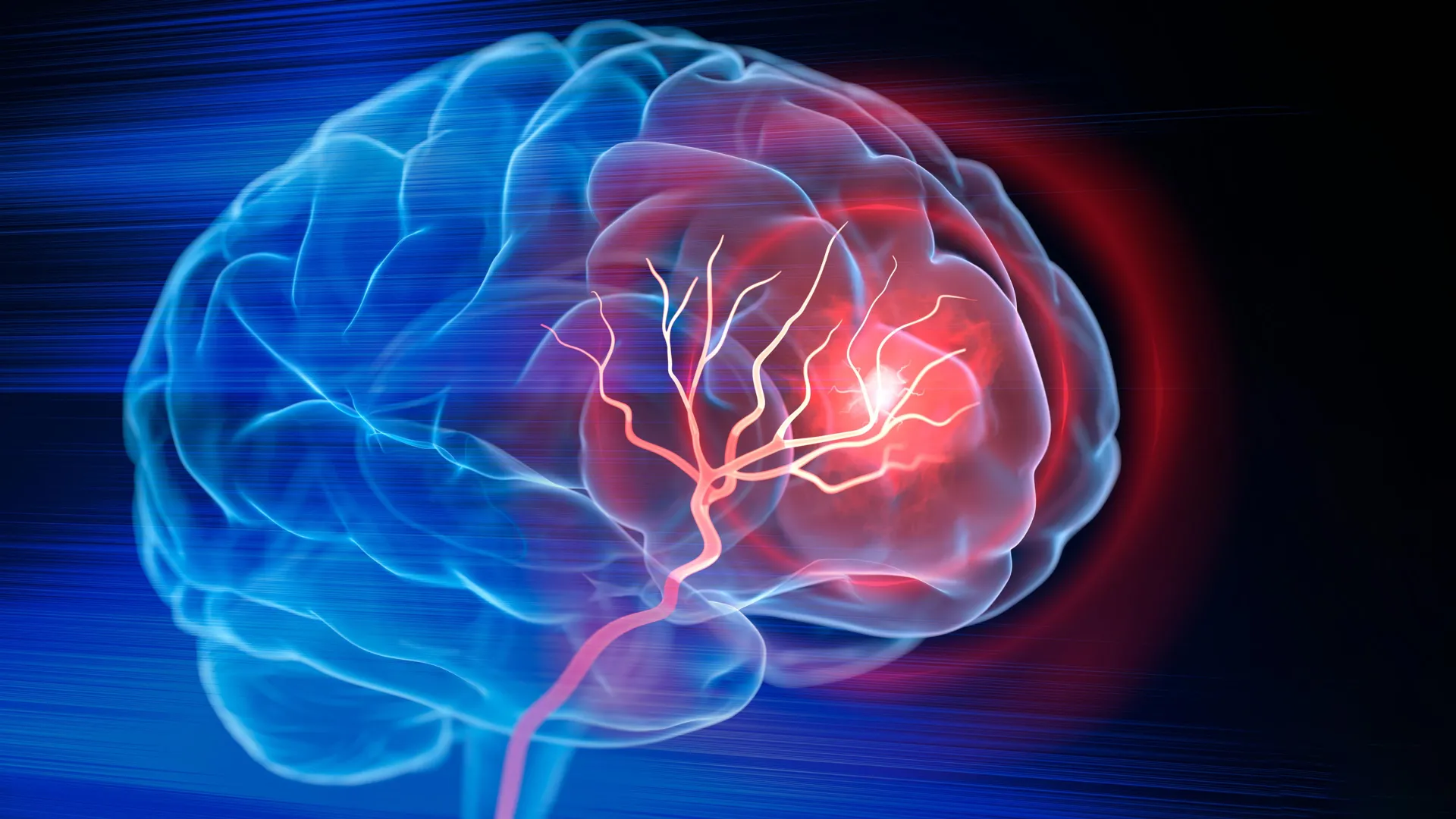
Orgo-Life the new way to the future Advertising by AdpathwayBefore birth, the human brain is assembled through an enormous series of cellular choices. At the center of this process are radial glia, a special type of stem cell that helps create many of the features that distinguish the human brain.
These cells produce large numbers of the neurons and support cells that make up the cerebral cortex, the brain region involved in thought, memory, and language. Radial glia are also believed to contribute to the unusually large expansion of the human cortex compared with that of other species. Although most disappear before birth, similar cells can later appear again in brain cancers for reasons scientists still do not fully understand.
"Radial glia are the coolest cells that have ever existed," said Aparna Bhaduri, an assistant professor of biological chemistry at the David Geffen School of Medicine at UCLA. "They're really key to making us human. But they're also at the center of many neurodevelopmental and neuropsychiatric disorders, as well as cancer -- so understanding how they make their decisions is one way to start understanding how those conditions arise."
Two new studies published in Cell and Science now provide a closer look at how radial glia make those developmental choices. Bhaduri and her colleagues found that the cells respond to two very different kinds of information: the way they process nutrients and direct physical signals from another part of the developing brain. Together, these findings offer new insight into how the human cortex produces its remarkable variety of cell types.
Metabolism helps direct brain stem cells
In the Cell study, researchers built a detailed map of metabolism in the developing human cortex. The project was a collaboration between Bhaduri's lab and Heather Christofk's lab and was led by co-first authors Jessenya Mil and Jose Soto.
To create the atlas, the team analyzed donated human tissue along with brain organoids grown from stem cells. Their results pointed to an unexpected conclusion: metabolism does not simply support brain development in the background. It can actively influence which kinds of cells are produced.
The researchers found that radial glia depend heavily on the pentose phosphate pathway, a metabolic process that uses glucose to make materials needed by cells that are dividing rapidly.
When the scientists lowered the amount of available glucose or interfered with this pathway, the stem cells changed what they produced. They began generating more inhibitory neurons and other cell types that normally appear later in development.
"What was surprising is that metabolism isn't just a passive thing that happens in the background," said Bhaduri, a member of both the UCLA Broad Stem Cell Research Center and the UCLA Health Jonsson Comprehensive Cancer Center. "It can really control how stem cells make decisions."
The results could help scientists investigate how maternal nutrition, metabolic disorders, and other environmental influences affect the developing brain. The metabolic atlas also provides one of the most detailed resources yet for researchers studying metabolism during human brain development.
A signal arrives early from the thalamus
The second study, published in Science and led by first author Claudia Nguyen, examined a completely different source of developmental information. This time, the researchers focused on signals coming from the thalamus, a structure deep inside the brain that helps relay information throughout the nervous system.
Scientists have known for years that neurons in the thalamus send long projections toward the cortex. These wire-like fibers eventually form connections with specific cortical neurons. However, anatomical studies have shown that in humans, the projections reach the cortex long before those final connections are established.
That raised an important question: Why do the fibers arrive so early?
Using human stem cell-derived brain "assembloids," the UCLA researchers found part of the answer. The thalamic projections physically touch radial glia while the brain is still developing.
That contact changed the behavior of the stem cells. It caused them to produce more excitatory neurons, the primary signal-carrying neurons in the cortex. The effect was especially strong for upper-layer neurons, which are particularly expanded in the human brain.
"We already knew that these projections influence how the cortex develops," Bhaduri said. "What we specifically found is that this influence comes through an actual physical connection between the projections and the radial glia -- a point of contact that just hasn't been identified before, and one that very likely does not exist in rodents."
A gene linked to autism enters the picture
The researchers connected this physical interaction to NRXN1, a gene already known for helping neurons form connections with one another. Mutations in NRXN1 have previously been associated with autism spectrum disorder.
To investigate its role, the team created assembloids from patient-derived cells carrying an NRXN1 mutation. In these models, the altered thalamic signals behaved differently from signals made by unaffected cells.
Those changes shifted the balance between the number of stem cells and the neurons they generated. The result offers researchers a possible way to study how disturbances early in brain development could influence the formation of the cortex.
The developing brain is in constant communication
Although the two studies focused on very different mechanisms, they point toward the same broader idea. One examined metabolism, while the other explored neural connections, yet both showed that radial glia do not make their decisions in isolation. Their behavior is continuously shaped by signals from the environment around them.
The studies also demonstrate how dramatically organoid technology has changed the study of human brain development. About a decade ago, scientists had few practical ways to directly investigate how uniquely human neural stem cells behave.
Today, brain organoids and related models allow researchers to recreate important features of human brain development in the laboratory. These systems also make it possible to test questions that cannot be addressed through animal models alone.
Bhaduri hopes the findings will encourage scientists to view metabolism and physical cellular connections as active drivers of development rather than as background processes.
"Ultimately, these studies give us a glimpse under the hood of how these cells make decisions," she said. "Understanding those decisions is a first step toward understanding normal brain development, disease vulnerability and, potentially, how similar stem-cell programs operate in brain cancer."
This research was supported by the National Institutes of Health, the National Science Foundation, the Brain & Behavior Research Foundation, the Alfred P. Sloan Foundation, the Rose Hills Foundation, the Esther A. & Joseph Klingenstein Fund, the Simons Foundation, the Chan Zuckerberg Initiative, the NIH BRAIN Initiative Cell Atlas Network, the International Foundation for Ethical Research, the UCLA Broad Stem Cell Research Center's Stem Cell Research Training Program, and the UCLA Health Jonsson Comprehensive Cancer Center and UCLA Broad Stem Cell Research Center Ablon Scholars Program.









.png)






.jpg)



 English (US) ·
  French (CA) ·