
 7 hours ago
1
7 hours ago
1
PROTECT YOUR DNA WITH QUANTUM TECHNOLOGY
Orgo-Life the new way to the future Advertising by AdpathwayFor the first time, researchers have directly seen and measured the protein clusters thought to spark Parkinson's disease, marking a major milestone in understanding the world's fastest-growing neurological condition.
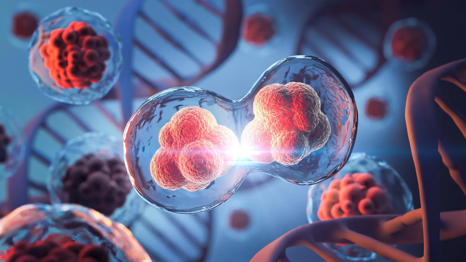
These microscopic clusters, known as alpha-synuclein oligomers, have long been suspected as the starting point for Parkinson's, but they have remained undetectable in human brain tissue -- until now.
A team from the University of Cambridge, UCL, the Francis Crick Institute, and Polytechnique Montréal developed a powerful imaging approach that allows scientists to visualize, count, and compare these protein clumps in human brain tissue. One researcher described the breakthrough as "like being able to see stars in broad daylight."
Published in Nature Biomedical Engineering, the findings could transform how scientists study Parkinson's, offering new insights into how it spreads through the brain and paving the way for earlier diagnosis and more targeted treatments.
Parkinson's: A Growing Global Health Challenge
More than 166,000 people in the UK currently live with Parkinson's disease, and the global total is expected to reach 25 million by 2050. While existing drugs can ease symptoms such as tremors and stiffness, none can halt or slow the disease's progression.
For over a century, doctors have identified Parkinson's by the presence of large protein deposits known as Lewy bodies. Yet researchers have long believed that smaller, early-stage oligomers may actually cause the damage to brain cells. Until now, these microscopic structures, just a few nanometers long, were impossible to observe directly.
Seeing Parkinson's at Its Earliest Stages
"Lewy bodies are the hallmark of Parkinson's, but they essentially tell you where the disease has been, not where it is right now," said Professor Steven Lee from Cambridge's Yusuf Hamied Department of Chemistry, who co-led the research. "If we can observe Parkinson's at its earliest stages, that would tell us a whole lot more about how the disease develops in the brain and how we might be able to treat it."
To achieve this, the researchers created a method called ASA-PD (Advanced Sensing of Aggregates for Parkinson's Disease). This ultra-sensitive fluorescence microscopy technique can detect and analyze millions of oligomers in post-mortem brain samples. Because the oligomers are so tiny, their signal is faint, but ASA-PD enhances that signal while reducing background noise, allowing scientists to clearly see individual alpha-synuclein clusters for the first time.
Illuminating the Invisible
"This is the first time we've been able to look at oligomers directly in human brain tissue at this scale: it's like being able to see stars in broad daylight," said co-first author Dr Rebecca Andrews, who conducted the work when she was a postdoctoral researcher in Lee's lab. "It opens new doors in Parkinson's research."
The researchers examined brain tissue from people with Parkinson's and compared it to samples from healthy individuals of similar age. They found that oligomers were present in both groups, but in those with Parkinson's, the clusters were larger, brighter, and far more numerous. This difference suggests a strong connection between oligomer growth and disease progression.
Clues to the Earliest Signs of Disease
The team also identified a unique subset of oligomers found only in Parkinson's patients, which may represent the earliest detectable signs of the disease -- possibly appearing years before symptoms emerge.
"This method doesn't just give us a snapshot," said Professor Lucien Weiss from Polytechnique Montréal, wo co-led the research. "It offers a whole atlas of protein changes across the brain and similar technologies could be applied to other neurodegenerative diseases like Alzheimer's and Huntington's.
"Oligomers have been the needle in the haystack, but now that we know where those needles are, it could help us target specific cell types in certain regions of the brain."
A New Window Into the Human Brain
"The only real way to understand what is happening in human disease is to study the human brain directly, but because of the brain's sheer complexity, this is very challenging," said Professor Sonia Gandhi from The Francis Crick Institute, who co-led the research. "We hope that breaking through this technological barrier will allow us to understand why, where and how protein clusters form and how this changes the brain environment and leads to disease."
This research was made possible with support from Aligning Science Across Parkinson's (ASAP), the Michael J. Fox Foundation, and the Medical Research Council (MRC), part of UK Research and Innovation (UKRI). The team expressed gratitude to the patients, families, and caregivers who donated brain tissue to research, enabling discoveries like this to advance understanding and potential treatment of Parkinson's disease.


.jpg)


 English (US) ·
  French (CA) ·